|
The Menisci (singular
form = meniscus) of the knee are crescent-shaped pads of tough,
rubbery fibrocartilage, which is a tissue commonly referred to
as "gristle" in the table meat processing industry.
The paired menisci of the human knee are often simply referred
to as the knee's "cartilages". They exist between the
femur (thigh bone) and tibia ("shin bone") to cushion
the knee joint during day-to-day use (see
FIGURES 1a-1c). Their specific job is to spread out the
joint's bone-to-bone contact pressure (caused by carrying your
body weight) over a broad area. This avoids concentrated stress
in any one spot, which can cause breakdown and deterioration (arthritis)
of the articular (gliding surface) cartilage covering the ends
of the femur and tibia. Several forms of meniscus damage and deterioration
are known to occur, which for general convenience have traditionally
been lumped together under the umbrella terms "meniscus tear"
or "torn cartilage".

FIGURE 1a - Basic knee anatomy, demonstrating the
location of the medial (inner) meniscus and lateral (outer)
meniscus. |
 FIGURE
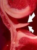
1b - This photograph demonstrates a cross-section of
the medial half of a normal human knee specimen. Above, you
see the rounded contour of the femoral condyle. Below, you
see the flatter, upper surface of the tibia. These two bones
have been drawn apart here to better demonstrate the meniscus
(see arrows), which lies between them. You can see that the
meniscus is a wedge-shaped structure when viewed in cross-section.
It is held in place here by its attachment to the knee's capsular
ligament on the left-hand side. FIGURE
1b - This photograph demonstrates a cross-section of
the medial half of a normal human knee specimen. Above, you
see the rounded contour of the femoral condyle. Below, you
see the flatter, upper surface of the tibia. These two bones
have been drawn apart here to better demonstrate the meniscus
(see arrows), which lies between them. You can see that the
meniscus is a wedge-shaped structure when viewed in cross-section.
It is held in place here by its attachment to the knee's capsular
ligament on the left-hand side. |
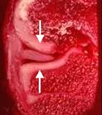 FIGURE
1c - In this picture, the same knee anatomy specimen
seen in Figure 1-b is shown, but in a more normal anatomic
configuration, with the femur (above) resting upon the tibia
(below). Here you can easily see how the meniscus serves as
a natural cushion or pad, interposed between the femur and
tibia. The arrows demonstrate the direction of joint loading
forces while standing. This specimen also demonstrates nicely
how the bones of the knee are lined with articular (joint
surface) cartilage (the white, border tissue coating the spongy-appearing,
dark red bone). Articular cartilage adds to the shock-absorbing
capability of the knee and provides the joint with smooth,
low-friction gliding surfaces. FIGURE
1c - In this picture, the same knee anatomy specimen
seen in Figure 1-b is shown, but in a more normal anatomic
configuration, with the femur (above) resting upon the tibia
(below). Here you can easily see how the meniscus serves as
a natural cushion or pad, interposed between the femur and
tibia. The arrows demonstrate the direction of joint loading
forces while standing. This specimen also demonstrates nicely
how the bones of the knee are lined with articular (joint
surface) cartilage (the white, border tissue coating the spongy-appearing,
dark red bone). Articular cartilage adds to the shock-absorbing
capability of the knee and provides the joint with smooth,
low-friction gliding surfaces. |
Just like the rest of our body parts, our menisci do age and
ultimately degenerate. While they can be suddenly torn apart by
a violent injury at any age, they typically become gradually weakened,
worn and broken down by natural processes as we get older, at
times causing symptoms while at other times not. The rearward
(posterior) portions of the menisci seem to receive the great
majority of stress, both in day-to-day life and in traumatic knee
sprains, thus almost all surgical work ends up being done on the
posterior two thirds of these structures. Tears of the frontal
(anterior) portions of the menisci occur only with relative rarity.
Because healthy menisci perform a useful function in the knee,
when injured they should be repaired and preserved whenever possible
and practical. Meniscus lesions are typically classified by orthopedic
surgeons as being either "traumatic" or "degenerative"
tears.
|